A new, quick and simple eye test can predict who is more at risk of age-related macular degeneration (AMD), a leading cause of blindness worldwide.
Researchers at Australia’s Vision Centre (VC) have found that while people with early AMD can still see in fine detail, other parts of their vision may be damaged and this isn’t revealed by current eye tests.
The new test, by revealing the damage, can show doctors or optometrists where to look and when to watch a patient more closely, helping them to lessen the risk of the disease and avoid total blindness.
“Many people as they grow older have little yellow blobs of protein and lipids that build up in the retina, known as drusen,” says Professor Ted Maddess of The VC and The Australian National University (ANU). “While having drusen doesn’t always lead to AMD, it can signal an increase in the individual’s risk of the eye disease.”
Prof. Maddess explains that AMD occurs when the drusen accumulate and expand, blocking the retina from getting oxygen or from disposing dead eye cell or tissue. This causes patches of the retina to die or leads to abnormal growth of blood vessels in the eye.
“It ends up damaging our central vision, which we use to read, drive and see in fine detail. This is why current tests for AMD focus on the central visual field,” he says. “However, these tests ignore other parts of the retina, which our study now shows can be damaged in early AMD.”
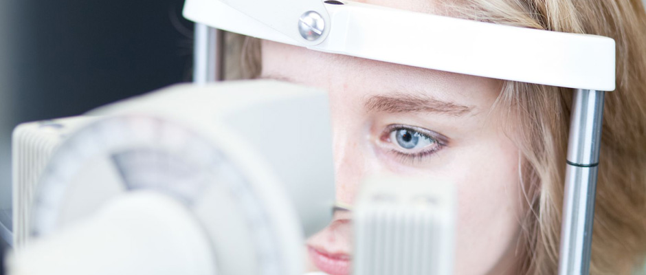
Using the TrueField Analyzer, a device developed by Prof. Maddess’s team and the Australian company “Seeing Machines”, Vision Centre researchers tested how pupils of early AMD respond to images on LCD screens. The images were provided to each eye at 44 locations in the person’s visual field.
Two video cameras using infrared lighting recorded the instantaneous response of the pupils, which was then analysed by a computer.
“All the patients had drusen and most could see in fine detail, which means that their central vision was still working well,” Prof. Maddess says. “However, when we measured other parts of their vision, we found that their pupils responded less compared to people without AMD.
“This shows that some of the drusen were harming parts of their vision, and the test reveals which patches were causing the damage.”
“While we don’t know if damage caused by drusen will always result in AMD, the test can act as a warning sign for doctors and patients,” Prof. Maddess says. “For instance, if there’s a patch of drusen near the central vision, and if the person’s vision is damaged in the same area, it’s a sign that they need close attention.
“Doctors can advise the patient to exercise more, to eat more antioxidants, or to stop habits like smoking that cause eye disease. They can also have the patient come in more frequently for check-ups.”
Prof. Maddess says AMD currently affects one in seven Australians over the age of 50, costs $2.6 billion a year, and will rob the central vision of 1.77 million Australians by 2030. In developed countries about 15 per cent of people over 40 show signs of early AMD, with about four per cent progressing to late stage AMD each year. “By the time eye tests detect damage in the central vision, it’s usually too late because it means the disease has progressed too far.
“Knowing who is at risk of progressing to late stage AMD and helping them to prevent it, regardless of whether they develop the disease or not, can go a long way towards saving their sight.”
The study “Multifocal pupillography identifies retinal dysfunction in early AMD” by Faran Sabeti, Andrew C. James, Rohan W. Essex and Ted Maddess was published in Graefe’s Archive for Clinical and Experimental Ophthalmology.
The Vision Centre is funded by the Australian Research Council as the ARC Centre of Excellence in Vision Science.