Conjunctivitis (also called pink eye or madras eye in India) is inflammation of the conjunctiva (the outermost layer of the eye and the inner surface of the eyelids). It is commonly due to an infection (usually viral, but sometimes bacterial) or an allergic reaction.
Signs and symptoms
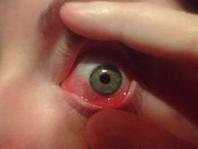
Red eye (hyperaemia), swelling of conjunctiva (chemosis) and watering (epiphora) of the eyes are symptoms common to all forms of conjunctivitis. However, the pupils should be normally reactive and the visual acuity normal.
Viral
Viral conjunctivitis is often associated with an infection of the upper respiratory tract, a common cold, and/or a sore throat. Its symptoms include excessive watering and itching. The infection usually begins with one eye, but may spread easily to the other.
Viral conjunctivitis, commonly known as pink eye, shows a fine, diffuse pinkness of the conjunctiva, which is easily mistaken for the ciliary injection of iritis, but there are usually corroborative signs on microscopy, particularly numerous lymphoid follicles on the tarsal conjunctiva, and sometimes a punctate keratitis.
Bacterial
Bacterial conjunctivitis causes the rapid onset of conjunctival redness, swelling of the eyelid, and mucopurulent discharge. Typically, symptoms develop first in one eye, but may spread to the other eye within 2–5 days. Bacterial conjunctivitis due to common pyogenic (pus-producing) bacteria causes marked grittiness/irritation and a stringy, opaque, greyish or yellowish mucopurulent discharge that may cause the lids to stick together, especially after sleep. Severe crusting of the infected eye and the surrounding skin may also occur, but, contrary to popular belief, discharge is not essential to the diagnosis. The gritty and/or scratchy feeling is sometimes localized enough for patients to insist they must have a foreign body in the eye. The more acute pyogenic infections can be painful. Common bacteria responsible for non-acute bacterial conjunctivitis are Staphylococci and Streptococci.
Bacteria such as Chlamydia trachomatis or Moraxella can cause a non-exudative but persistent conjunctivitis without much redness. Bacterial conjunctivitis may cause the production of membranes or pseudomembranes that cover the conjunctiva. Pseudomembranes consist of a combination of inflammatory cells and exudates, and are loosely adherent to the conjunctiva, while true membranes are more tightly adherent and cannot be easily peeled away. Cases of bacterial conjunctivitis that involve the production of membranes or pseudomembranes are associated with Neisseria gonorrhoeae, β-hemolytic streptococci, and C. diphtheriae. Corynebacterium diphtheriae causes membrane formation in conjunctiva of non immunized children.
Chemical
Chemical eye injury is due to either an acidic or alkali substance getting in the eye. Alkalis are typically worse than acidic burns.Mild burns will produce conjunctivitis while more severe burns may cause the cornea to turn white. Litmus paperis an easy way to rule out the diagnosis by verifying that the pH is within the normal range of 7.0—7.2. Large volumes of irrigation is the treatment of choice and should continue until the pH is 6—8.Local anaesthetic eye drops can be used to decrease the pain.
Irritant or toxic conjunctivitis show primarily marked redness. If due to splash injury, it is often present only in the lower conjunctival sac. With some chemicals, above all with caustic alkalis such as sodium hydroxide, there may be necrosis of the conjunctiva with a deceptively white eye due to vascular closure, followed by sloughing of the dead epithelium. This is likely to be associated with slit-lamp evidence of anterior uveitis.
Other
Inclusion conjunctivitis of the newborn (ICN) is a conjunctivitis that may be caused by the bacteria Chlamydia trachomatis, and may lead to acute, purulent conjunctivitis.However, it is usually self-healing.
Conjunctivitis is identified by irritation and redness of the conjunctiva. Except in obvious pyogenic or toxic/chemical conjunctivitis, a slit lamp (biomicroscope) is needed to have any confidence in the diagnosis. Examination of the tarsal conjunctiva is usually more diagnostic than the bulbar conjunctiva.